Facelift surgery began as an operation that primarily tightened skin and evolved into surgery that anatomically mobilizes and repositions deeper facial tissues. Although many procedure names are used today, the differences become easier to understand when we ask which layer is dissected, how far the dissection extends and how the tissues are managed.
The beginning of facelift surgery
The first documented facelifts were performed in the early twentieth century. In 1901, Eugen von Holländer reportedly removed excess skin and closed the defect with little or no undermining. Later procedures extended the operation by excising skin around the ear and dissecting beneath the skin before redraping it.
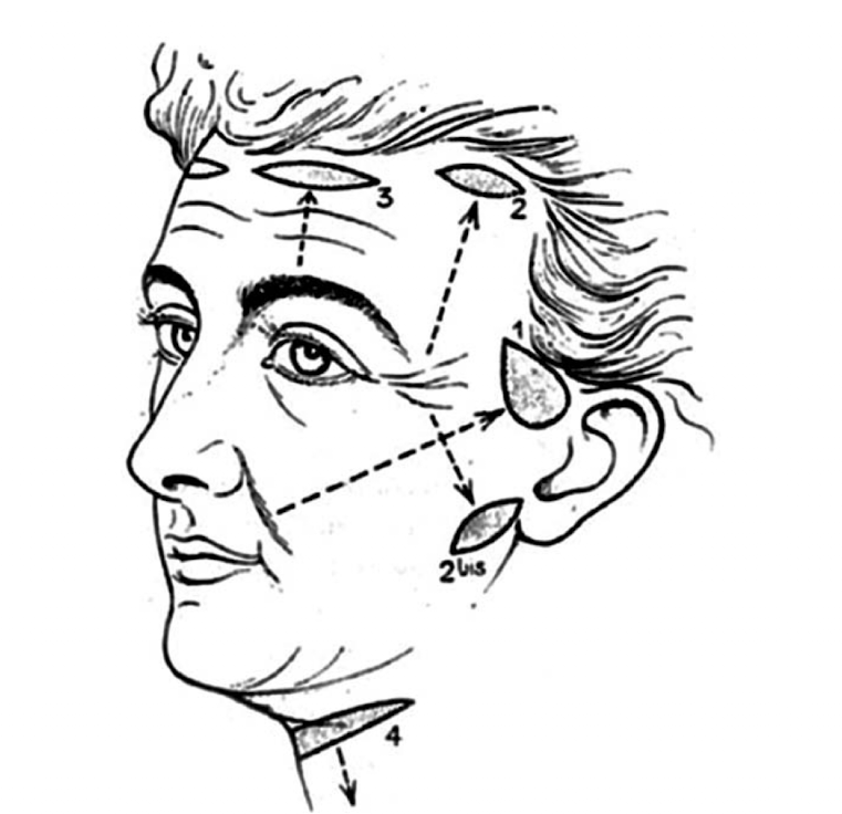
The principal tissue in these early operations was the skin. An immediate tightening effect was possible, but relying on stretchable skin created several limitations: recurrence could occur, excessive tension could be transferred to the scar, and deeper laxity of the cheek and jawline was difficult to reposition effectively.
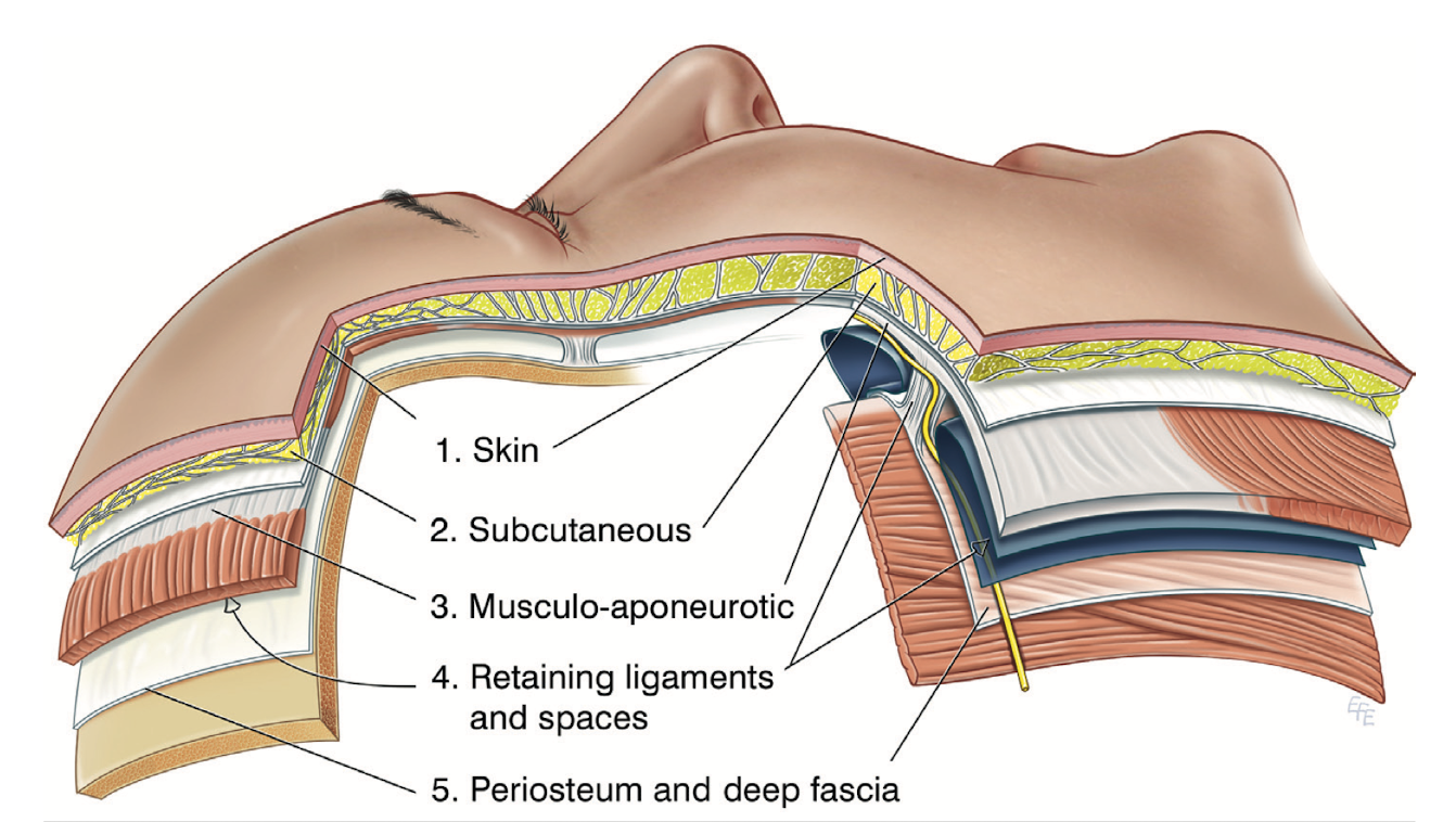
The five facial layers
The facial soft tissues can be understood in five layers from superficial to deep: skin, subcutaneous tissue, the SMAS and its continuous structures, facial spaces, and periosteum or deep fascia. Modern facelift techniques differ in the plane of dissection, the retaining structures released and the tissues mobilized.
Moving deeper: Skoog and the SMAS
In the late 1960s, Swedish plastic surgeon Tord Skoog described lifting not only the skin but also the superficial fascia of the face and the platysma of the neck as a continuous unit. His approach, later systematized in a 1974 text, helped establish the foundation for modern facelifts that work beneath the skin.
In 1976, Mitz and Peyronie described the superficial musculoaponeurotic system (SMAS) through anatomical studies of the parotid and cheek regions. Once surgeons had this shared anatomical language, multiple techniques emerged: separating skin from deeper tissues, folding or excising the SMAS, and repositioning it in different directions.
Excess skin was removed and the lift relied primarily on the skin.
Skin, superficial fascia and platysma were treated as a continuous unit, shifting attention to deeper tissues.
Mitz and Peyronie described the SMAS anatomically, leading to a wider variety of techniques for deeper tissue management.
Three categories of modern facelift surgery
Modern facelifts carry many names. Here, representative techniques are organized into three groups according to the relationship between the skin and SMAS and the depth at which dissection is performed.
This is not the only possible classification. Surgeons may use the same name for operations with different dissection or fixation, and some procedures combine more than one concept. The categories are best viewed as a map for understanding the operation.
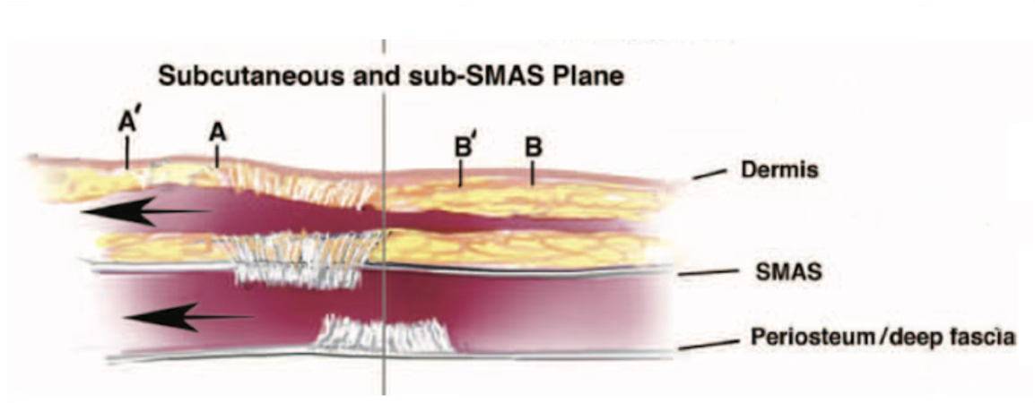
Skin and SMAS are mobilized separately
Examples: High SMAS / Extended SMAS
The skin is dissected and the SMAS is then mobilized as a separate layer. Because each layer can be moved with a different vector and degree of tension, deeper tissue repositioning can be balanced against tension on the skin.
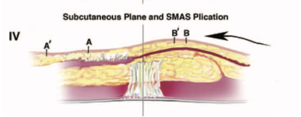
Subcutaneous dissection with surface-level SMAS manipulation
Examples: SMAS plication / SMASectomy / MACS lift
Dissection is performed mainly beneath the skin. Without wide sub-SMAS dissection, the SMAS may be folded, tightened with sutures, partially excised and repaired, or suspended. This group generally permits a more limited operative field than techniques involving broad sub-SMAS dissection.
Sub-SMAS dissection with composite tissue movement
Examples: Deep Plane / Composite Facelift
The operation enters beneath the SMAS and releases the retaining structures required for mobilization. Skin, subcutaneous fat and SMAS are moved as a continuous, thicker unit.
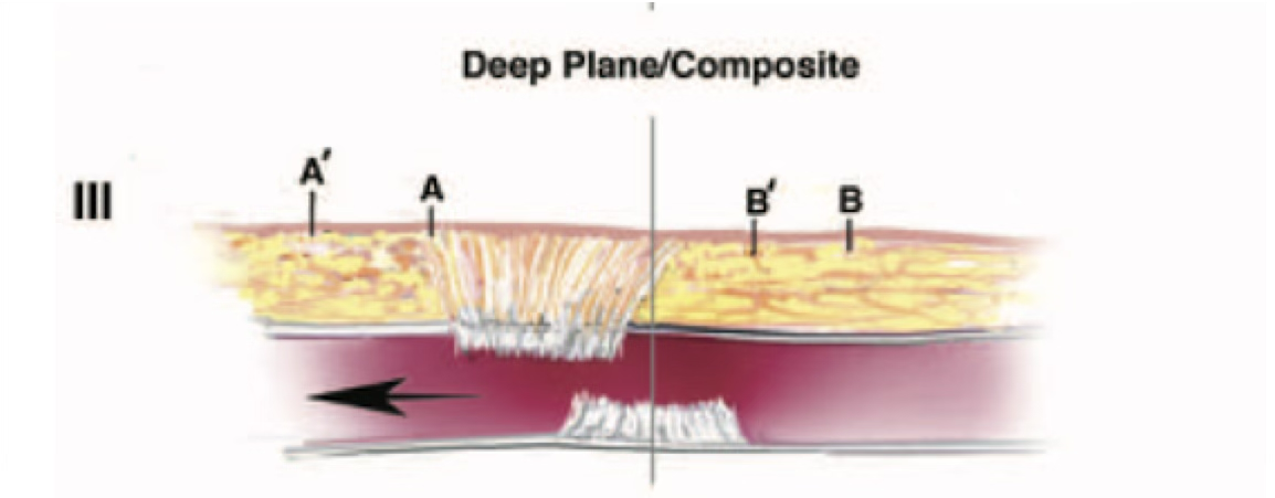
What the three categories tell us
These categories describe more than a difference in names. Whether the skin and deeper tissues are mobilized separately, the SMAS is managed from its surface, or the dissection enters beneath the SMAS and moves the tissues together affects the dissection field, the way tissues move, patterns of swelling and recovery, relevant anatomy and the resulting contour.
Deeper is not automatically better, and a smaller operative field is not automatically safer in every patient. The appropriate technique depends on the location and degree of laxity, the condition of the skin, fat and SMAS, previous surgery and the change the patient hopes to achieve.
Three questions for understanding a technique
- What is the main plane of dissection: subcutaneous, sub-SMAS, or both?
- How is the SMAS managed?
- Which retaining structures are released, how far are they released, and how are the tissues moved?
Summary
The history of facelift surgery can be viewed as an evolution from skin-only tightening to anatomically guided repositioning of deeper tissues that include the SMAS.
Today’s many techniques become easier to compare through three questions: whether the skin and SMAS are separated, how the SMAS is managed, and how extensively the retaining structures are released. The actual surgical layer and extent matter more than the procedure name alone.